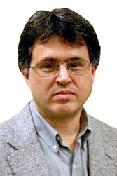
Dr. Ayrton Bernussi and Dr. Luis Grave de Peralta Awarded $345,000 NSF Grant to Develop a Versatile Microscope Digital LED Condenser
By Amanda Miller


Development of high-resolution and improved technology microscopes has allowed scientists and engineers to achieve images of structures with greater quality than was previously possible, including the ability to see structures never seen before. Numerous industries have taken advantage of this technology, from biomedical engineering to chip manufacturing. These various applications for microscopes have led to many new techniques, such as dark field microscopy, phase-contrast microscopy, and ptychography — all using different technology or image analysis to yield different results. However, currently, these diverse techniques with their unique applications all require slightly different microscope light sources or condenser designs, and therefore a separate individual microscope.
Texas Tech University's Dr. Luis Grave de Peralta, associate professor of physics, and Dr. Ayrton Bernussi, associate professor of electrical and computer engineering, are conducting research that will impact the microscopy world through the development of a new, versatile, one-part microscope digital condenser comprised of individually and electronically controlled LEDs. This new condenser will operate on a revolutionary technology — doubling resolution, increasing contrast, and allowing multiple microscopy techniques on one device. The duo's hope is that the digital condenser may be — once tested and optimized — produced for adoption in high schools and universities, and even in the many industries — which widely rely on microscopy for new breakthroughs in the understanding of common manufacturing processes.

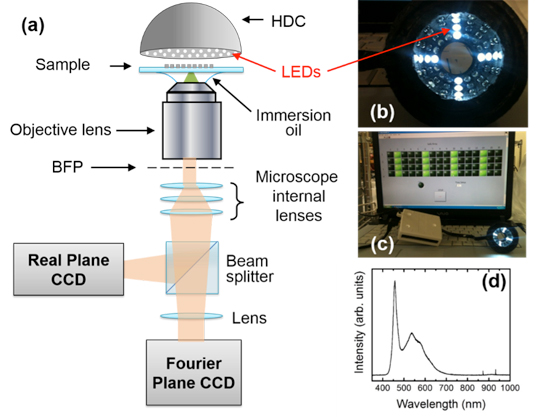
(b) Photograph of the HDC
(c) Associated electronic and computer
(d) Emission spectrum of the LEDs
(S. Sen et. al. , Biomedical Optics Express 6, 658-667 (2015).
Traditional microscopes operate using a single light source for illuminating the sample, lenses or mirrors, and several moving parts (iris, diaphragm, etc.). This translates into a more complicated device and limits the abilities of any one microscope, as it can only have one type of light as its source. In contrast, Grave de Peralta and Bernussi are creating a new condenser that is the shape of a hollow hemisphere made of plastic and is created using a 3D printer. This hemisphere is lined with anywhere from 64 to 460 electronically and individually controlled LEDs. Each LED can be turned on at different times, can be comprised of different colors, and can be used in different combinations with others inside the same condenser. It is possible to illuminate an object, therefore, by turning on a single LED, a combination of LEDs, row of these lights, etc. It is also possible to use different types of LEDs in order to yield different images. For example, Grave de Peralta and Bernussi have already successfully created a condenser using white light LEDs and are testing its use on various samples. But, it is possible to incorporate infrared as well as ultraviolet LEDs in combination with visible light in order to provide a single microscope with various capabilities. With a $346,663 grant from the National Science Foundation (NSF), the two will develop a condenser equipped with infrared LEDs for many profound applications.
Infrared microscopy is widely utilized in the semiconductor manufacturing industries, where silicon structures are unable to be seen utilizing visible light. These structures are very small and are encased in layers of material on a wafer. The wafers appear black to the naked eye and defects in these processed structures can only be imaged utilizing infrared microscopes. The condenser that will be developed utilizing this grant will be capable of accomplishing this task. In addition, because the condensers can be made using a combination of diverse LEDs, there is potential for them to be also adopted by and have important implications for biological science, the biomedical industry, metrology and inspection, and even the optoelectronic industry. For example, biological samples absorb light at different frequencies; therefore, using multiple LEDs that can be controlled and used in a number of combinations could allow higher-quality images in medicine than was possible before.
Sanchari Sen, a graduate student studying electrical engineering and physics, is helping to develop samples and image analysis techniques for the new condensers. Because of her extensive hands-on experience with this research, she has been offered a summer internship with Intel. There are also several other master's students involved in this project. It is Grave de Peralta's and Bernussi's end goal to make new and improved imaging possible for a more versatile microscope than conventional microscopes of the past. These devices are less expensive and require less power than previous microscope components. Therefore, the revolutionary condensers will save money and energy while promoting higher-capability microscopy for countless applications.

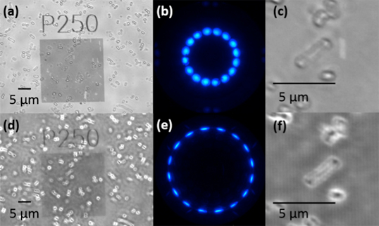
(b) snd (e) Fourier Plane (FP) images of E. Coli bacteria obtained using the HDC.
(c) and (f) were obtained by zooming in the RP images shown in (a) and (d), respectively.
(S. Sen et. al. , Biomedical Optics Express 6, 658-667 (2015).
Grant Title:
Edward E. Whitacre Jr. College of Engineering
-
Address
100 Engineering Center Box 43103 Lubbock, Texas 79409-3103 -
Phone
806.742.3451 -
Email
webmaster.coe@ttu.edu
